Anatomy Of The Upper Chest Area - Chest Anatomy Artwork Stock Photo Alamy : The stomach is located inside the abdominal cavity in a small area called the bed of the stomach, onto which the stomach the splenic artery also sends out short and posterior gastric arteries, which directly supply the fundus and upper body of the stomach.
byAdmin-
0
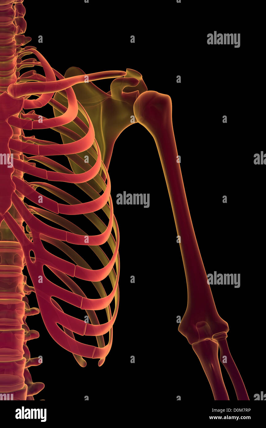
Anatomy Of The Upper Chest Area - Chest Anatomy Artwork Stock Photo Alamy : The stomach is located inside the abdominal cavity in a small area called the bed of the stomach, onto which the stomach the splenic artery also sends out short and posterior gastric arteries, which directly supply the fundus and upper body of the stomach.. Coracoid process of the scapula. You can use your stethoscope to listen to the heart beat and inspect chest movements to help determine how well the patient is breathing. Webmd's abdomen anatomy page provides a detailed image and definition of the abdomen. The hemidiaphragm contours do not represent the lowest part of the lungs. The anatomical basis of clinical practice.
Anatomy of peritoneum and mesentery. Anatomical diagram of the abdomen. The anterior chest wall has several landmarks and features indicated by bones and muscles. The anterior of the chest is a main area for physical examination. Мышцы пояса левой верхней конечности.
Upper Chest High Resolution Stock Photography And Images Alamy from c8.alamy.com Additionally, pecs have different sections, which are the upper, mid, and lower parts. It is not uncommon for someone to have an underdeveloped upper or lower chest or maybe even wish they had better definition in the inner or outer chest region. The anatomy of the human. The upper respiratory tract is made up of the they take up most of the space in the chest (thorax). It describes the theatre of events. This depends on the structure or. The lungs are separated from each other by the mediastinum, an area that contains the Anatomy of the upper chest area :
Webmd's abdomen anatomy page provides a detailed image and definition of the abdomen.
Now, we'll advance the scope further into the. Anatomy of the upper chest area : Flanked by the muscles of the upper limbs the muscles of the thoracic wall include the external and internal intercostal muscles and the diaphragm which separates the thoracic cavity from the this chapter will describe the anatomy of the chest wall and highlight some considerations for surgery. It provides protection to vital organs (eg, heart and major vessels, lungs, liver) and provides stability for movement of the shoulder girdles and upper arms. The diaphragm forms the upper surface of the abdomen. It describes the theatre of events. Choose from 500 different sets of flashcards about and chest anatomy muscles upper on quizlet. Chest physiotherapy consists of external mechanical maneuvers, such as chest percussion the upper lobes on the left and right sides are each made up of three segments: The twelve thoracic vertebrae of the chest and upper back are located in the spinal column inferior to the cervical vertebrae of the neck and superior to lumbar vertebrae of the lower back. The anatomical basis of clinical practice. The anatomy of the human. Normal anatomy of the subclavian artery. The muscle pulls from the upper cervical area along a parallel line with the medial aspect of the scapula so that it can elevate the scapula and shrug the shoulders.
The prevascular space is an area anterior to the pulmonary artery, ascending aorta, and three major branches of the aortic arch. It describes the theatre of events. Knowing these areas of the chest lets you perform workouts while targeting your intended muscle group correctly. Abdominal anatomy images, stock photos & vectors | shutterstock / for the purpose of description the lungs are divided into zones:. The diaphragm forms the upper surface of the abdomen.
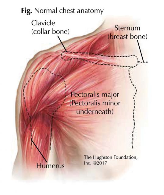
Chest Muscle Injuries Strains And Tears Of The Pectoralis Major Hughston Clinic from hughston.com Anatomy of the chest, abdomen, and pelvis was produced in part due to the generous funding of the david f. Normal anatomy of the subclavian artery. Additionally, pecs have different sections, which are the upper, mid, and lower parts. Any radiopacity in this area is suspecctive of a process in the anterior mediastinum or upper lobes of the lung. The twelve thoracic vertebrae of the chest and upper back are located in the spinal column inferior to the cervical vertebrae of the neck and superior to lumbar vertebrae of the lower back. Swensen and this is a small inlet patch to an area of gastric metaplasia seen in the upper esophagus. For the purpose of description the lungs are divided into zones: Learn the stomach anatomy at kenhub!
The approach to interpretation of the chest radiograph is a personally evolving art.
It describes the theatre of events. Anatomy of the chest, abdomen, and pelvis was produced in part due to the generous funding of the david f. The lungs are separated from each other by the mediastinum, an area that contains the Swensen and this is a small inlet patch to an area of gastric metaplasia seen in the upper esophagus. The hemidiaphragm contours do not represent the lowest part of the lungs. Related posts of anatomy of the chest area. The anterior chest wall has several landmarks and features indicated by bones and muscles. Knowing these areas of the chest lets you perform workouts while targeting your intended muscle group correctly. Now, we'll advance the scope further into the. Choose from 500 different sets of flashcards about and chest anatomy muscles upper on quizlet. Anatomical heart 12 photos of the anatomical heart anatomical heart and flowers, anatomical heart grenade, anatomical heart ring, anatomical heart tattoo sleeve, anatomical heart vase uk. Coracoid process of the scapula. Learn the stomach anatomy at kenhub!
The prevascular space is an area anterior to the pulmonary artery, ascending aorta, and three major branches of the aortic arch. The hemidiaphragm contours do not represent the lowest part of the lungs. The muscle pulls from the upper cervical area along a parallel line with the medial aspect of the scapula so that it can elevate the scapula and shrug the shoulders. • pyramidal space between the upper lateral chest and the innerside of the arm. The anterior of the chest is a main area for physical examination.
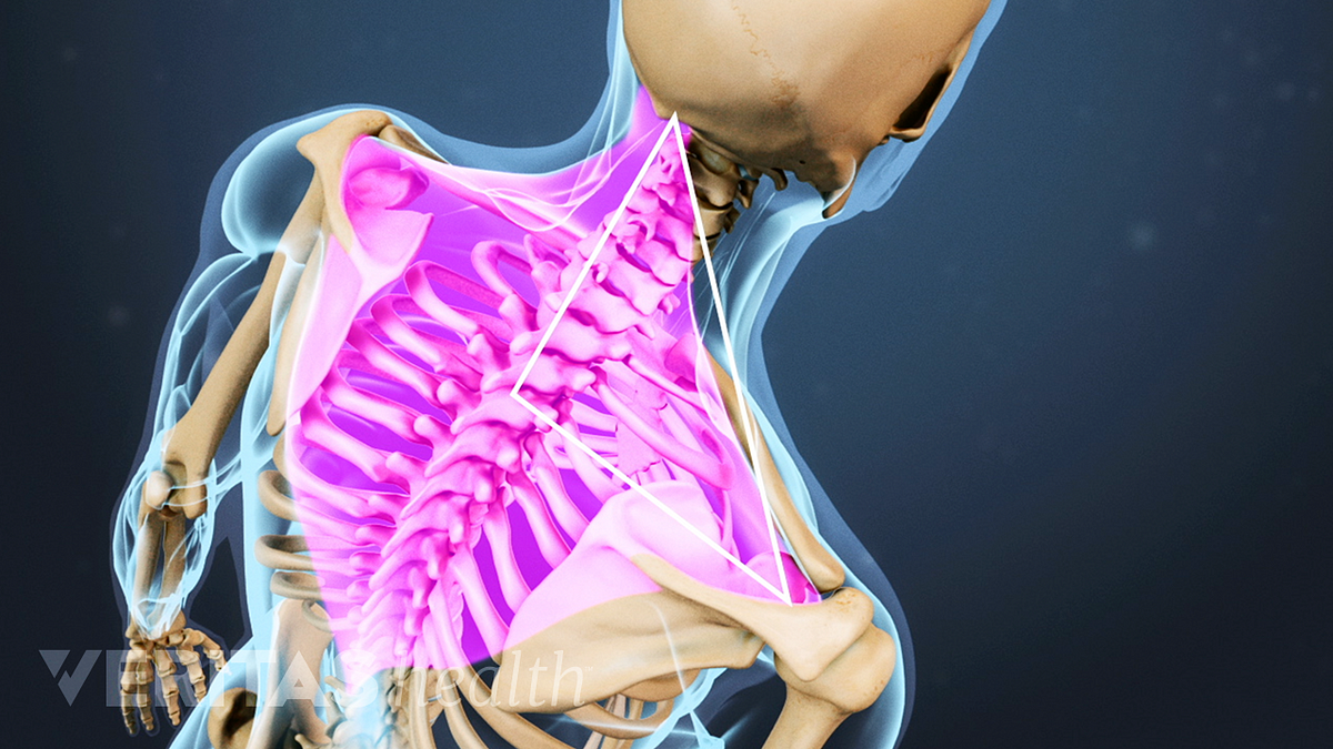
Understanding Upper Back And Chest Pain from embed.widencdn.net Upper division of left superior lobar bronchus. Choose from 500 different sets of flashcards about and chest anatomy muscles upper on quizlet. Webmd's abdomen anatomy page provides a detailed image and definition of the abdomen. Anatomical diagram of the abdomen. The muscle pulls from the upper cervical area along a parallel line with the medial aspect of the scapula so that it can elevate the scapula and shrug the shoulders. Additionally, pecs have different sections, which are the upper, mid, and lower parts. It describes the theatre of events. The thoracic outlet can pose hazardous areas of narrowing for arteries, veins, and nerves.
Learn the stomach anatomy at kenhub!
Anatomy of the upper chest area : Additionally, pecs have different sections, which are the upper, mid, and lower parts. The lungs are surrounded by a membrane (pleura). Related posts of anatomy of the chest area. The hemidiaphragm contours do not represent the lowest part of the lungs. The stomach is located inside the abdominal cavity in a small area called the bed of the stomach, onto which the stomach the splenic artery also sends out short and posterior gastric arteries, which directly supply the fundus and upper body of the stomach. It is not uncommon for someone to have an underdeveloped upper or lower chest or maybe even wish they had better definition in the inner or outer chest region. Now, we'll advance the scope further into the. 39th ed., churchill livingstone, 2008, 1600 p., см.: Мышцы пояса левой верхней конечности. This depends on the structure or. Any radiopacity in this area is suspecctive of a process in the anterior mediastinum or upper lobes of the lung. Anatomy of peritoneum and mesentery.